"Open the Door and Let Me In"
Building a Model of a Cell Membrane

Background:
The cell membrane regulates what enters and leaves the cell and aids in the protection and support of the cell. In many ways the cell membrane can be compared to the walls that surround a house. As these walls help to protect the house from what is outside, so does the membrane seal off the cell from its outside environment. But if you lived in the house, you would still want to receive messages, fuel, and power from outside. You would also need to bring in food and take out trash. Thus, doors would be needed. The needs of the cell are similar; it must communicate with other cells, take in food and water, and eliminate wastes. All of this is possible because of the structure of the cell membrane.
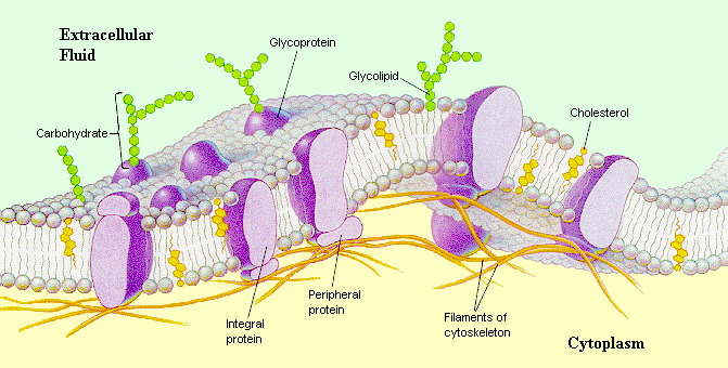
The cell membrane is a layer about nine nanometers (nine billionths of a meter) thick, covered with pores, proteins, and twiglike carbohydrate groups that project from some of the proteins and lipids. The membrane is constructed of molecules called phospholipids, whose chemical structure forms two layers. The hydrophobic (water hating) tails face each other in the middle of the membrane, much like the buttered sides of the bread in a sandwich. The hydrophilic (water loving) heads face the watery environment on each side of the membrane. This structure gives the membrane fluidity to allow selective permeability (passage of certain materials but not others).
Some proteins stick to the surface of the lipid bilayer, whereas others are free to move around within the layer. Many of the free-moving ones act as channels through which molecules pass. Some of the proteins control the passage of water-soluble substances into and out of cells, acting like small pumps, actively pushing molecules from one side of the membrane to the other, much like a revolving door. Still others form enzymes that participate in metabolic reactions. Structural proteins, such as microfilaments, bind with the plasma membrane to maintain a cell's shape and stability.
The carbohydrate projections are believed to function as surface receptors that gather chemical messages; as markers that help cells identify each other; as control points to regulate cell growth. Cholesterol is found only in animal cells and is located between the phospholipid molecules. It increases the flexibility of the membrane.

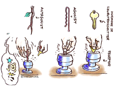
How receptor sites work and how drugs affect them
Aim: To make a three-dimensional model of a cell membrane.
Materials: paper fasteners or cereal (Kix™), string, assorted pasta, marshmallows, pony beads, pipe cleaners, glue, construction paper
Procedure:
1. Construct the bilipid layer of the cell membrane using paper fasteners or Kix™ cereal and string. Glue to illustrate the thickness of the membrane.
2. Glue other shapes to show the protein and carbohydrate molecules. (Refer to the Introduction and the diagram.)
3. Construct a key for your model.
Follow-Up
1. Place the following words in your glossary:
|
phagocytosis |
pinocytosis |
diffusion |
|
|
osmosis |
hypotonic |
hypertonic |
|
|
Isotonic |
exocytosis |
endocytosis |
|
2. In what way is a plasma membrane selective?
3. Using a Venn diagram, compare and contrast active and passive transport.
